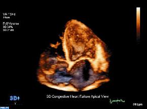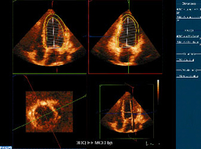
3-D Echocardiography
Technique:
A 3-D ultrasound transducer (wand-like apparatus) is placed on the chest to produce the clearest image of the heart. The state-of-the-art 3-dimensional transducer, advanced software, and high-definition imaging system is capable of acquiring a virtual image of the entire heart that is able to be manipulated in 3-dimensional space. This enables Dr. Scuderi to view every structure contained within the heart from any plane producing test results that are far more refined than traditional 2-dimensional imaging. This world-class technology is available to you at CardioFit and yields results with the highest sensitivity and specificity so that your heart is thoroughly evaluated and the most accurate diagnosis is made possible.
Purpose:
To check how well the heart is moving, the size of the heart, and its pumping chambers (ventricles).
Preparation:
The test will take 30-45 minutes depending on the patient’s condition and the type of echocardiogram needed.
Benefits of 3-D echocardiographic imaging:
Live 3-D imaging
- Real-time, 360 degree visualization of cardiac structures and cardiac blood flow
- Precise measurements superior to cardiac magnetic resonance imaging (MRI) regarding assessment of left ventricular (LV) volume, right ventricular (RV) volume, and hypertrophic cardiomyopathy
- Rivals cardiac MRI regarding measurements of LV systolic function, LV volume, LV mass, left atrial volume, ASD, VSD, as well as aortic valve and mitral valve area
- Automatically calculates LV ejection fraction resulting in highly reproducible measurements
- Markedly reduces the need for invasive testing